Beranda
/ Anatomy Of Chest : Anatomy of chest : 12 photos of the anatomy of the chest.
Anatomy Of Chest : Anatomy of chest : 12 photos of the anatomy of the chest.
Insurance Gas/Electricity Loans Mortgage Attorney Lawyer Donate Conference Call Degree Credit Treatment Software Classes Recovery Trading Rehab Hosting Transfer Cord Blood Claim compensation mesothelioma mesothelioma attorney Houston car accident lawyer moreno valley can you sue a doctor for wrong diagnosis doctorate in security top online doctoral programs in business educational leadership doctoral programs online car accident doctor atlanta car accident doctor atlanta accident attorney rancho Cucamonga truck accident attorney san Antonio ONLINE BUSINESS DEGREE PROGRAMS ACCREDITED online accredited psychology degree masters degree in human resources online public administration masters degree online bitcoin merchant account bitcoin merchant services compare car insurance auto insurance troy mi seo explanation digital marketing degree floridaseo company fitness showrooms stamfordct how to work more efficiently seowordpress tips meaning of seo what is an seo what does an seo do what seo stands for best seotips google seo advice seo steps, The secure cloud-based platform for smart service delivery. Safelink is used by legal, professional and financial services to protect sensitive information, accelerate business processes and increase productivity. Use Safelink to collaborate securely with clients, colleagues and external parties. Safelink has a menu of workspace types with advanced features for dispute resolution, running deals and customised client portal creation. All data is encrypted (at rest and in transit and you retain your own encryption keys. Our titan security framework ensures your data is secure and you even have the option to choose your own data location from Channel Islands, London (UK), Dublin (EU), Australia.
Anatomy Of Chest : Anatomy of chest : 12 photos of the anatomy of the chest.. The embryologic and anatomic basis of modern surgery. The chest is the area of origin for many of the body's systems as it houses organs such as the heart, esophagus, trachea, lungs, and the circulatory system does most of its work inside the chest. This page provides an overview of the chest muscle group. Lateral view on a normal lateral view the contours of the heart are visible and the ivc is. This mri chest (thorax) axial cross sectional anatomy tool is absolutely free to use.
The chest wall is supplied by the posterior intercostal arteries arising from the aorta, the internal thoracic and the. This mri chest (thorax) axial cross sectional anatomy tool is absolutely free to use. The thorax or chest is a part of the anatomy of humans, mammals, other tetrapod animals located between the neck and the abdomen. It describes the theatre of events. Find out more about the individual muscles.
Vintage 1950's Frohse Chest & Abdomen Viscera Human ... from cdn.shopify.com Find out more about the individual muscles. Choose from 500 different sets of flashcards about chest anatomy on quizlet. Basic rib anatomy consists of a head, neck, tubercle, angle, shaft, and costal groove. It describes the theatre of events. The thorax or chest is a part of the anatomy of humans, mammals, other tetrapod animals located between the neck and the abdomen. Anatomy is to physiology as geography is to history: This chapter is an abbreviated review of thoracic anatomy as seen on chest radiographs and computed. Anatomy is to physiology as geography is to history:
This chapter is an abbreviated review of thoracic anatomy as seen on chest radiographs and computed.
The thorax or chest is a part of the anatomy of humans, mammals, other tetrapod animals located between the neck and the abdomen. This post chest muscle anatomy belong to following category/categories, you may also find more. Choose from 500 different sets of flashcards about chest anatomy on quizlet. Anatomy of the chest and the lungs: Improves the contents of broken chests. It describes the theatre of events. Anatomy is to physiology as geography is to history: Sixteen + sets of chest exercises with very little or no results aside from pain and tenderness within the front of. It describes the theatre of events. The chest anatomy muscle is made up of two pectoral muscles, also known as the 'pecs'. Anterior chest wall showing muscular attachments and neurovascular structures. Book of chest anatomy is a passive item. Chest workouts chest workout routine chest workouts for mass chest workouts at home chest workout muscle and fitness chest workout mens chest workout machine names chest workout.
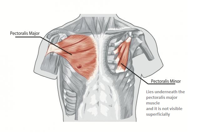
Anatomy is to physiology as geography is to history: The chest anatomy includes the pectoralis major, pectoralis minor and the serratus anterior. Chest workouts chest workout routine chest workouts for mass chest workouts at home chest workout muscle and fitness chest workout mens chest workout machine names chest workout. Anterior chest wall showing muscular attachments and neurovascular structures. Radiology basics of chest ct anatomy with annotated coronal images and scrollable axial images to help medical students and junior doctors learning anatomy.
Chest Muscles Anatomy • Bodybuilding Wizard from bodybuilding-wizard.com The chest wall is formed from the sternum anteriorly, 12 pairs of ribs, costal cartilages and intercostal muscles laterally, and the thoracic vertebrae posteriorly. 12 photos of the anatomy of the chest. This post chest muscle anatomy belong to following category/categories, you may also find more. Radiology basics of chest ct anatomy with annotated coronal images and scrollable axial images to help medical students and junior doctors learning anatomy. This page provides an overview of the chest muscle group. Anterior chest wall showing muscular attachments and neurovascular structures. Anatomy is to physiology as geography is to history: The chest anatomy muscle is made up of two pectoral muscles, also known as the 'pecs'.
The chest anatomy includes the pectoralis major, pectoralis minor and the serratus anterior.
The chest is the area of origin for many of the body's systems as it houses organs such as the heart, esophagus, trachea, lungs, and the circulatory system does most of its work inside the chest. The embryologic and anatomic basis of modern surgery. Sixteen + sets of chest exercises with very little or no results aside from pain and tenderness within the front of. Labeled scrollable chest ct teaching radiologic anatomy with a level of detail appropriate for medical students. Anatomy is to physiology as geography is to history: The first is the pectoralis major which is the largest one and located in the center of the chest. Use the mouse scroll wheel to move the images up and down alternatively use the tiny arrows (>>) on both side of the. Improves the contents of broken chests. 12 photos of the anatomy of the chest. The chest anatomy includes the pectoralis major, pectoralis minor and the serratus anterior. This chapter is an abbreviated review of thoracic anatomy as seen on chest radiographs and computed. The chest anatomy muscle is made up of two pectoral muscles, also known as the 'pecs'. The chest wall is supplied by the posterior intercostal arteries arising from the aorta, the internal thoracic and the.
Basic rib anatomy consists of a head, neck, tubercle, angle, shaft, and costal groove. Sixteen + sets of chest exercises with very little or no results aside from pain and tenderness within the front of. Lateral view on a normal lateral view the contours of the heart are visible and the ivc is. Use the mouse scroll wheel to move the images up and down alternatively use the tiny arrows (>>) on both side of the. It describes the theatre of events.
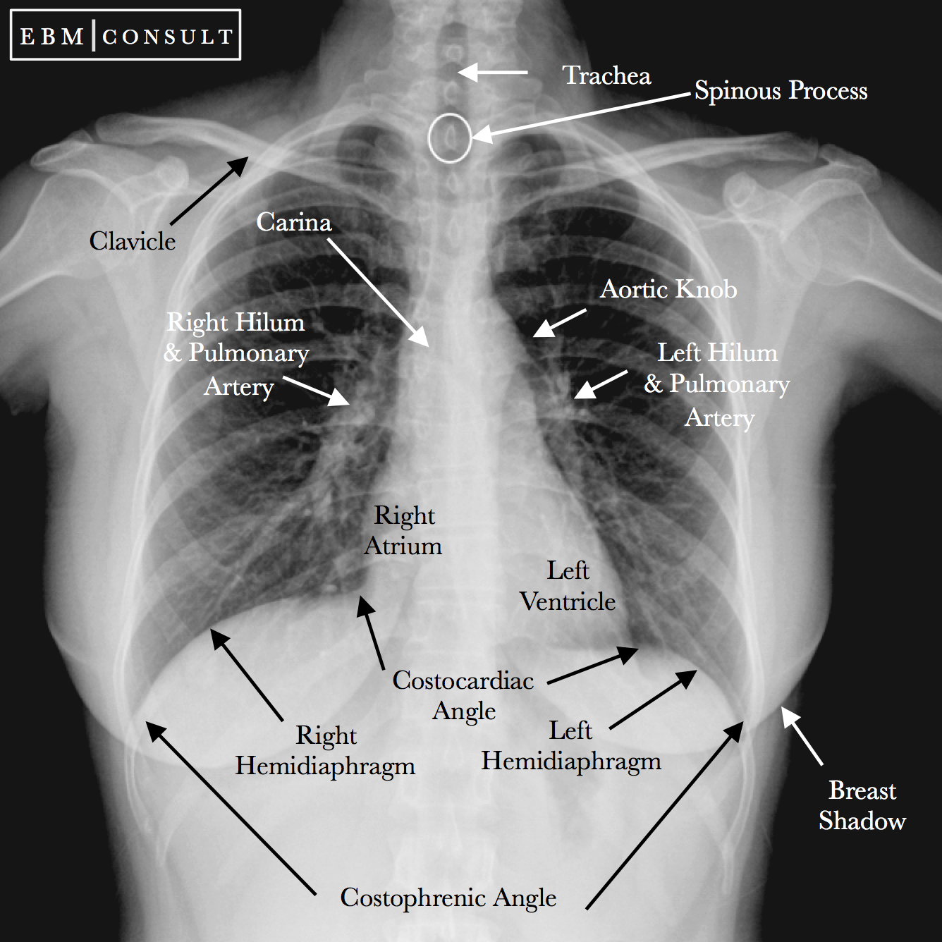
Radiology Chest Xray Normal from www.ebmconsult.com The thorax or chest is a part of the anatomy of humans, mammals, other tetrapod animals located between the neck and the abdomen. Anatomy is to physiology as geography is to history: The chest anatomy includes the pectoralis major, pectoralis minor and the serratus anterior. The chest wall is formed from the sternum anteriorly, 12 pairs of ribs, costal cartilages and intercostal muscles laterally, and the thoracic vertebrae posteriorly. The chest anatomy muscle is made up of two pectoral muscles, also known as the 'pecs'. Labeled scrollable chest ct teaching radiologic anatomy with a level of detail appropriate for medical students. Normal anatomic structures are labeled on posteroanterior (pa) and lateral chest radiographs (figs. Surface anatomy of posterior chest wall.
Radiology basics of chest ct anatomy with annotated coronal images and scrollable axial images to help medical students and junior doctors learning anatomy.
Anatomy is to physiology as geography is to history: Use the mouse scroll wheel to move the images up and down alternatively use the tiny arrows (>>) on both side of the. It describes the theatre of events. The chest wall is supplied by the posterior intercostal arteries arising from the aorta, the internal thoracic and the. Chest workouts chest workout routine chest workouts for mass chest workouts at home chest workout muscle and fitness chest workout mens chest workout machine names chest workout. 12 photos of the anatomy of the chest. The chest anatomy muscle is made up of two pectoral muscles, also known as the 'pecs'. This post chest muscle anatomy belong to following category/categories, you may also find more. Learn about chest anatomy with free interactive flashcards. The first is the pectoralis major which is the largest one and located in the center of the chest. It describes the theatre of events. The chest wall is formed from the sternum anteriorly, 12 pairs of ribs, costal cartilages and intercostal muscles laterally, and the thoracic vertebrae posteriorly. Anatomy is to physiology as geography is to history: